History and Biology of Streptococcus pneumoniae

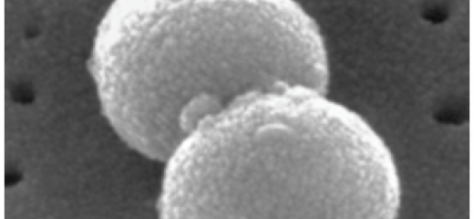
Streptococcus pneumoniae are lancet-shaped (elongated round shape) Gram-positive bacteria that can either grow in chains or pairs as shown in this image. It was first isolated independently in 1880 in France by Louis Pasteur from the saliva of a patient who had rabies and in the USA by George M. Sternberg.
In 1886, S.pneumoniae was first known as pneumococcus due to its close association with pneumonia. In 1920, it was renamed as Diplococcus pneumoniae as it was usually observed in pairs. In 1974, the name was changed to Streptococcus pneumoniae due to its similarities in forming chains like other Streptococcus spp. S. pneumoniae can be distinguished from other species using rMLST.
S.pneumoniae is a leading global cause of morbidity and mortality amongst all age groups but children and the elderly are most at risk. The burden of pneumococcal disease is highest in low- and middle-income countries. S.pneumoniae causes mild infections like otitis media and sinusitis, but also life-threatening infections like pneumonia, septicaemia and meningitis. Worldwide, pneumococcal disease is highest among children less than 5 years of age and an estimated 1.6 million deaths occur every year, mainly due to pneumonia.

Streptococcus pneumoniae seen by electron micrograph
Biology
Factors that enable pneumococci to evade the immune system and cause disease include the polysaccharide capsule, various surface proteins, and toxins such as the pneumolysin gene (ply).
The polysaccharide capsule is a key virulence factor because it protects the pneumococcus and enables it to evade phagocytosis by the host immune system.
There are at least 100 antigenically different polysaccharide capsules or ‘serotypes’. The capsule is the target for the current pneumococcal vaccines. Pneumolysin is a toxin that forms pores in the cell membrane and causes cell lysis, which induces inflammation and activates the complement system in the human host.
S.pneumoniae can undergo phase variation, allowing it to have different phenotypes during colonisation and invasion. During colonisation, S.pneumoniae can produce a thin (or transparent) capsule, better adapted for colonisation, whereas during invasion a thicker (opaque) capsule is observed, which enables it to evade phagocytosis.

Another factor that enables bacteria to survive is that pneumococci can change their capsule to avoid being recognised by the immune system. They do this by taking up DNA (transformation) encoding a different set of capsular genes from another pneumococcus and incorporating that new DNA into their genome (recombination), discarding the original capsular genes. This process of transformation and recombination also enables the pneumococcus to acquire other advantageous genes like antibiotic-resistance genes.
History - Epidemics
S.pneumoniae causes disease in all countries around the world. Outbreaks are possible but not as large scale or as frequent as meningitis outbreaks caused by Neisseria meningitidis. Seasonal outbreaks of bacterial meningitis due to S.pneumoniae have been recorded in the meningitis belt. Most of these cases were predominantly due to serotype 1 pneumococci, which is especially prevalent in Africa.
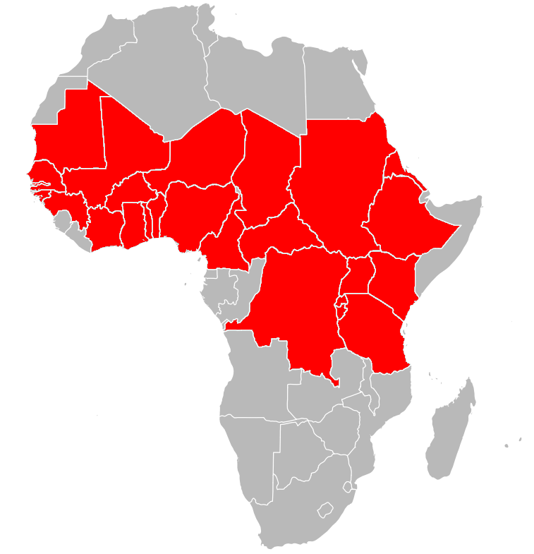
Meningitis outbreaks highlight the need for current and continuous revision of epidemic prevention, surveillance and diagnostic strategies, which to date have largely focused on outbreaks caused by N.meningitidis.
To learn more about Streptococcus pneumoniae click on the links bellow.